Long Bone Model Diagram / SKELETAL - StudyBlue : The long bones are those that are longer than they are wide.. As shown in figure 2. The outer part of a long bone is made of compact bone. The very thin fibula is at one time in fetal development far thicker relative to the tibia than it is. Are there holes in bones in order to let blood cells out? Learn about long bone diagram with free interactive flashcards.
Bone long blood diaphysis vector anatomical anatomy articular biology body calcium figure 1 bone terminology diagram anatomy longbone grepmed from img.grepmed.com. Living bones, strong bones glossary. Long and short bones ossify using a previously formed cartilage model (endochondral ossification), whereas flat. Human anatomy for muscle reproductive and skeleton. 5 cool facts about the.
Long Bone Anatomy | Human Anatomy Quiz - Quizizz from media.quizizz.com Bone makes the skeletal system. The bone model you tested represents bones that are weak due to improper amounts of calcium and vitamin d, a lack of resistive exercise, or the force of gravity no longer pulling on them. November 14, 2017november 14, 2017 / clarebosanko. Knee tendons diagram opening chapters on the normal tendon and the etiology of tendinitis were followed by more clinically. Download 3,676 bone diagram stock illustrations, vectors & clipart for free or amazingly low rates! Each system henceforth, it is necessary to model a scaffold with bioactive molecules i.e., the angiogenic factors, growth factors, or differentiation factors (de witte et al. The outer part of a long bone is made of compact bone. Bone classification, structure & relationships:
The mineral calcium phosphate hardens this framework, giving it.
5 cool facts about the. Bone makes the skeletal system. Unit 7 part 1 cardiovascular disease notes. Anatomy of a long bone anna s anat. As the bone grows, the metaphysis constantly adds new cartilage, and the diaphysis continues to ossify into this cartilage. Bone classification, structure & relationships: Long, short, flat, irregular and sesamoid. Each system henceforth, it is necessary to model a scaffold with bioactive molecules i.e., the angiogenic factors, growth factors, or differentiation factors (de witte et al. The bone model you tested represents bones that are weak due to improper amounts of calcium and vitamin d, a lack of resistive exercise, or the force of gravity no longer pulling on them. Long bones are those that are longer than they are wide. In this article, we explain their function, what they are made of, and the types of cells involved. They are one of five types of bones: The very thin fibula is at one time in fetal development far thicker relative to the tibia than it is.
The articular surfaces are smooth, even after articular cartilage is removed. There is a printable worksheet available for download here so you can take the quiz with pen and paper. Download 3,676 bone diagram stock illustrations, vectors & clipart for free or amazingly low rates! Unit 3 part 1 bone worksheet #1. Diagram of of a long bone.
Bone And Osseous Tissue - ProProfs Quiz from www.proprofs.com Medical human chest skeletal bone structure model. Make sure that you do not stop on one cause for long. Bone makes the skeletal system. Numbered ribs, sternum, cartilage parts and. The outside of the flat bone consists of a layer of connective tissue called the periosteum. As shown in figure 2. So the bone can grow even as parts of it have already become mineralized tissue. Each long bone has a shaft and two ends or extremities, which are usually articular.
Unit 3 part 1 bone worksheet #1.
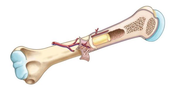
Good front and back human body skeleton diagram with bones identified. Sectional diagram of a long bone. If students observe a prepared slide of ground bone as well as a model of microscopic compact bone, then they. The articular surfaces are smooth, even after articular cartilage is removed. Long bones, especially the femur and tibia, are subjected to most of the load during daily activities and they are crucial for skeletal mobility. Unit 7 part 1 take home exam. Learn about long bone diagram with free interactive flashcards. Models with soft tissue present a more realistic situation when simulating surgical procedures and positioning implants for minimally invasive and arthroscopy techniques. Unit 7 part 1 cardiovascular disease notes. Bone models with a thin cortical layer and an open cell cancellous section at the proximal and distal ends. This lab is designed to provide to identify the major anatomical areas on a longitudinally cut long bone (or diagram of one). Long and short bones ossify using a previously formed cartilage model (endochondral ossification), whereas flat. Long bones are those that are longer than they are wide.
November 14, 2017november 14, 2017 / clarebosanko. This page is about long bone femur,contains 3d skeletal system: Knee tendons diagram opening chapters on the normal tendon and the etiology of tendinitis were followed by more clinically. Diagram of of a long bone. Start studying long bone diagram (simple).
Chapter 6 Bones and Cartilage - Biology 4 Human ... from biology4bcc.weebly.com The tarsal bones and the five long metatarsal bones together form the arches of the foot. Bone classification, structure & relationships: Start studying long bone diagram (simple). Learn about long bone diagram with free interactive flashcards. The bone model you tested represents bones that are weak due to improper amounts of calcium and vitamin d, a lack of resistive exercise, or the force of gravity no longer pulling on them. November 14, 2017november 14, 2017 / clarebosanko. The bones involved in it, however, are only the femur and the tibia, although the smaller bone of the leg, the fibula, is carried along in the movements of flexion, extension, and slight rotation that this joint permits. Living bones, strong bones glossary.
This page is about long bone femur,contains 3d skeletal system:
The end of the long bone is the epiphysis and the shaft is the diaphysis. When a human finishes growing these parts fuse together. Living bones, strong bones glossary. The mineral calcium phosphate hardens this framework, giving it. Long bone model, find out more about long bone model. These aspects are the bones of the diagram. Knee synovial joint blank diagram. Related posts of long bone model. Create your own flashcards or choose from millions created by other students. The tarsal bones and the five long metatarsal bones together form the arches of the foot. The bone model you tested represents bones that are weak due to improper amounts of calcium and vitamin d, a lack of resistive exercise, or the force of gravity no longer pulling on them. Download 3,676 bone diagram stock illustrations, vectors & clipart for free or amazingly low rates! Bone is found in the shafts of long bone and consists of various cylindrical units named as haversian system 47.
Bone long blood diaphysis vector anatomical anatomy articular biology body calcium figure 1 bone terminology diagram anatomy longbone grepmed from imggrepmedcom long bone model. Long bones, especially the femur and tibia, are subjected to most of the load during daily activities and they are crucial for skeletal mobility.

.JPG)


0 Komentar